Structure and Role of Different Parts of the Eye
Introduction
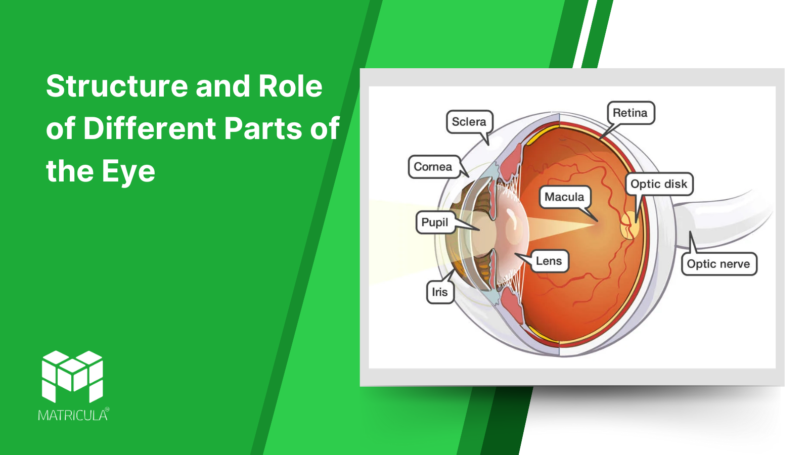
The human eye is a highly specialized sense organ that plays a crucial role in vision. It functions by detecting light, converting it into electrical impulses, and transmitting these signals to the brain via the optic nerve. The ability to perceive colors, shapes, and depth depends on the intricate coordination between different parts of the eye. Each part has a specific structure and function that contributes to clear and accurate vision.
In this detailed guide, we will explore the different parts of the eye, including the sclera, cornea, choroid, iris, retina, aqueous humour, lens, vitreous humour, conjunctiva, eyelids, and tear glands—their structure, function, and how they contribute to visual perception.
1. Sclera (The White of the Eye)
Structure:
- The sclera is the outermost protective layer of the eyeball.
- It is composed of dense connective tissue and appears white in color.
- It extends from the cornea in the front to the optic nerve at the back of the eye.
Role:
- Provides structural support and maintains the shape of the eyeball.
- Protects the internal delicate structures of the eye from injury and external damage.
- Serves as an attachment point for muscles that control eye movement.
2. Cornea (The Transparent Window of the Eye)
Structure:
- The cornea is the clear, dome-shaped outermost layer covering the front of the eye.
- It lacks blood vessels but receives oxygen and nutrients from the aqueous humour and tear film.
- It has five layers, including the epithelium, stroma, and endothelium, which maintain its transparency and function.
Role:
- Acts as the primary refractive surface, bending light rays to focus them onto the retina.
- Protects the eye from dust, germs, and harmful particles.
- Works with the lens to focus light for clear vision.
3. Choroid (The Vascular Layer of the Eye)
Structure:
- The choroid is a thin, pigmented layer located between the sclera and retina.
- It contains numerous blood vessels that nourish the retina and other parts of the eye.
- Its dark pigmentation reduces internal light reflection, improving visual clarity.
Role:
- Provides oxygen and nutrients to the retina and other eye tissues.
- Absorbs excess light, preventing blurred vision caused by internal reflections.
- Plays a role in thermoregulation, maintaining a stable temperature for proper retinal function.
4. Iris (The Colored Part of the Eye)
Structure:
- The iris is a circular, pigmented muscle that determines eye color.
- It contains two types of muscles:
- The circular muscles (sphincter pupillae) that constrict the pupil.
- The radial muscles (dilator pupillae) that dilate the pupil.
Role:
- Controls the amount of light entering the eye by adjusting the pupil’s size.
- In bright light, the pupil constricts to reduce light entry.
- In dim light, the pupil dilates to allow more light in, improving vision in darkness.
5. Retina (The Light-Sensitive Layer)
Structure:
- The retina is the innermost layer of the eye, composed of photoreceptor cells.
- Contains two types of photoreceptors:
- Rods (responsible for black-and-white vision and night vision).
- Cones (responsible for color vision and sharp details).
- The fovea centralis, a small central part of the retina, has a high concentration of cones, providing sharp vision.
Role:
- Converts light into electrical signals, which are transmitted to the brain through the optic nerve.
- The brain processes these signals, enabling the perception of images.
- Helps in differentiating colors, shapes, and brightness.
6. Aqueous Humour (The Fluid of the Eye)
Structure:
- A transparent, watery fluid found in the anterior chamber between the cornea and the lens.
- Continuously produced by the ciliary body.
Role:
- Maintains intraocular pressure, keeping the eyeball firm.
- Provides oxygen and nutrients to the cornea and lens.
- Removes metabolic waste from eye tissues.
7. Lens (The Natural Optical Lens of the Eye)
Structure:
- A biconvex, transparent structure located behind the iris.
- It is held in place by suspensory ligaments attached to the ciliary muscles.
Role:
- Adjusts its shape to focus light on the retina, a process called accommodation.
- Helps in sharp near and distant vision.
- Works with the cornea to refract light accurately.
8. Vitreous Humour (The Gel-like Substance of the Eye)
Structure:
- A clear, gel-like substance filling the space between the lens and retina.
Role:
- Maintains the shape of the eyeball.
- Allows light to pass through to the retina without distortion.
- Provides support to the retina.
9. Conjunctiva (The Protective Membrane of the Eye)
Structure:
- A thin, transparent mucous membrane covering the sclera and lining the inner eyelids.
Role:
- Protects the eye from dust, microbes, and irritants.
- Keeps the eye moist and lubricated.
- Plays a role in immune defense by preventing infections.
10. Eyelids (The Protective Cover of the Eye)
Structure:
- Composed of skin, muscle, and glands, designed to open and close over the eye.
Role:
- Protects the eye from injury, bright light, and debris.
- Spreads tear fluid evenly over the cornea.
- Prevents dryness by reducing tear evaporation.
11. Tear Glands (Lacrimal Glands – The Moisture Provider)
Structure:
- Small, almond-shaped glands located above the outer corners of the eyes.
Role:
- Produce tears, which keep the eye moist and lubricated.
- Help in removing foreign particles and preventing infections.
- Contain antibacterial enzymes that protect against microbes.
The eye is a highly coordinated organ where different structures work together to ensure clear and accurate vision. The cornea and lens focus light, the iris regulates light entry, the retina captures images, and the optic nerve sends signals to the brain. Protective structures like the sclera, conjunctiva, eyelids, and tear glands ensure that the eye remains healthy.
Understanding these components is essential for appreciating how vision works and maintaining good eye health. Regular eye check-ups and proper care can help prevent vision-related disorders.